
Page 71 - 2024-bfw-MyersAP4e
P. 71
Module 1.4b
Structure of the Cortex
If you opened a human skull, exposing the brain, you would see a wrinkled organ, shaped
somewhat like an oversized walnut. Without these wrinkles, a flattened cerebral cortex
would require triple the area — roughly that of a large pizza. The brain’s left and right hemi-
spheres are filled mainly with axons connecting the cortex to the brain’s other regions. The
cerebral cortex — that thin surface layer — contains some 20 to 23 billion of the brain’s nerve
cells and 300 trillion synaptic connections (de Courten-Myers, 2005). Being human takes a
lot of nerve.
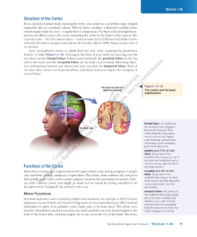
Each hemisphere’s cortex is subdivided into four lobes, separated by prominent
fissures, or folds (Figure 1.4-12). Starting at the front of your brain and moving over the
Distributed by Bedford, Freeman & Worth Publishers. Not for redistribution.
top, there are the frontal lobes (behind your forehead), the parietal lobes (at the top
and to the rear), and the occipital lobes (at the back of your head). Reversing direc-
tion and moving forward, just above your ears, you find the temporal lobes. Each of
the four lobes carries out many functions, and many functions require the interplay of
Copyright © Bedford, Freeman & Worth Publishers.
several lobes.
Figure 1.4-12
The brain has left and
right hemispheres The cortex and its basic
subdivisions
Frontal lobe Parietal lobe
Temporal lobe
frontal lobes the portion of
Occipital lobe the cerebral cortex lying just
behind the forehead. They
enable linguistic processing,
muscle movements, higher-
order thinking, and executive
functioning (such as making
plans and judgments).
parietal [puh-RYE-uh-tuhl]
lobes the portion of the
cerebral cortex lying at the top of
the head and toward the rear; it
receives sensory input for touch
Functions of the Cortex and body position.
More than a century ago, surgeons found damaged cortical areas during autopsies of people occipital [ahk-SIP-uh-tuhl]
who had been partially paralyzed or speechless. This rather crude evidence did not prove lobes the portion of the
that specific parts of the cortex control complex functions like movement or speech. A lap- cerebral cortex lying at the back
of the head; it includes areas that
top with a broken power cord might go dead, but we would be fooling ourselves if we receive information from the
thought we had “localized” the internet in the cord. visual fields.
temporal lobes the portion of
Motor Functions the cerebral cortex lying roughly
Scientists had better luck in localizing simpler brain functions. For example, in 1870, German above the ears; it includes the
physicians Gustav Fritsch and Eduard Hitzig made an important discovery: Mild electrical auditory areas, each of which
receives information primarily
stimulation to parts of an animal’s cortex made parts of its body move. The effects were from the opposite ear. They also
selective: Stimulation caused movement only when applied to an arch-shaped region at the enable language processing.
back of the frontal lobe, running roughly ear-to-ear across the top of the brain. Moreover,
The Brain: Brain Regions and Structures Module 1.4b 71
03_myersAPpsychology4e_28116_ch01_002_163.indd 71 15/12/23 9:23 AM