
Page 60 - 2022-bfw-morris-1e
P. 60
The third piece of information came from Jerry a. b.
Donohue and John Griffith, colleagues of Watson and Crick 3’ 5’ 3’ 5’
at Cambridge University. They determined that if the bases Sugar– Sugar–
phosphate
©2022 BFW Publishers. PAGES NOT FINAL. For review purposes only - do not post.
were to pair in some way, the most likely way would be that phosphate backbone
backbone
A paired with T and that G paired with C.
An accurate model of DNA had to account for the Bases
results of all of these pieces of information. Watson and Bases
Crick went to work, using sheet metal cutouts of the bases
and wire ties for the sugar–phosphate backbone. After many
false starts, they finally found a structure that worked: a
double- helical structure with the backbones on the outside,
the bases pointing inward, and A paired with T and G paired
with C.
The two scientists realized immediately that they had
made one of the most important discoveries in all of biology.
That day, February 28, 1953, they lunched at the Eagle, a
pub across the street from their laboratory, where Crick
loudly pronounced, “We have discovered the secret of life.”
The Eagle is still there in Cambridge, England, and on its
wall is a commemorative plaque marking the table where
the two ate. Watson and Crick published the structure of
DNA in 1953. Knowing the structure of DNA opened the
door to understanding how genetic information is stored,
faithfully replicated, and able to direct the synthesis of other
macromolecules.
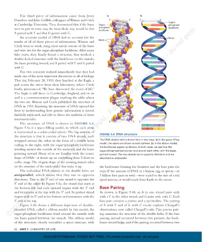
The structure of DNA is shown in FIGURE 5.6 . 3’ 5’ 3’ 5’
Figure 5.6a is a space-filling model, in which each atom T G C A
is represented as a color-coded sphere. The big surprise of
the structure is that it consists of two DNA strands, each FIGURE 5.6 DNA structure
wrapped around the other in the form of a double helix The DNA double helix is shown here in two ways. (a) In the space-filling
coiling to the right, with the sugar–phosphate backbones model, the atoms are shown as solid spheres. (b) In the ribbon model,
winding around the outside of the molecule and the bases the backbones appear as ribbons. In both cases, we see how the
sugar–phosphate backbones twist around each other, with the bases
pointing inward. Many of us are familiar with the iconic pointed inward. The two strands run in opposite directions and are
shape of DNA—it shows up on everything from T-shirts to described as antiparallel.
coffee mugs. The elegant shape of the twisting strands relies
on the structure of the nucleotides that make it up. the backbones forming the banisters and the base pairs the
The individual DNA strands in the double helix are steps. If the amount of DNA in a human egg or sperm—or
antiparallel , which means that they run in opposite 3 billion base pairs in total—were scaled to the size of a real
directions. That is, the 3′ end of one strand is opposite the spiral staircase, it would reach from Earth to the moon.
5′ end of the other. In Figure 5.6a, the strand that starts at
the bottom left and coils upward begins with the 3′ end Base Pairing
and terminates at the top with the 5′ end. Its partner strand As shown in Figure 5.6b, an A in one strand pairs only
begins with its 5′ end at the bottom and terminates with the with a T in the other strand, and G pairs only with C. Each
3′ end at the top. base pair contains a purine and a pyrimidine. The pairing
Figure 5.6b shows a different depiction of double- of A with T and of G with C nicely explains Chargaff ’s
stranded DNA, called a ribbon model. In this model, the observations, now called Chargaff’s rule. This precise pair-
sugar–phosphate backbones wind around the outside with ing maintains the structure of the double helix. If the base
the bases paired between the strands. The ribbon model pairing instead occurred between two purines, the back-
of the structure closely resembles a spiral staircase, with bones would bulge, and if the pairing occurred between two
82 UNIT 1 cHeMisTRY OF liFe
©2022 BFW Publishers. PAGES NOT FINAL. For review purposes only - do not post.
08_morrisapbiology1e_11331_Unit1_Mod5_78-91_2pp.indd 82 30/03/21 9:54 AM