
Page 128 - 2024-bfw-MyersAP4e
P. 128
Unlike cones, rods congregate in the retina’s outer regions. Rods remain sensitive in
dim light, and they enable black-and-white vision. Rods have no hotline to the brain. If
cones are soloists, rods perform as a chorus. Several rods pool their faint energy output and
funnel it onto a single bipolar cell, which sends the combined message to your brain.
Cones and rods each provide a special sensitivity — cones to detail and color, and
rods to faint light and peripheral motion. Stop for a minute and experience this rod–cone
difference. Pick a word in this sentence and stare directly at it, focusing its image on the
cones in your fovea. Notice that words distant from it appear blurred? Their image is strik-
ing your retina’s outer regions, where rods predominate. Thus, when you drive or bike,
rods help you detect a car in your peripheral vision well before you perceive its details.
How many of the black dots can you see at once in Figure 1.6-11?
Distributed by Bedford, Freeman & Worth Publishers. Not for redistribution.
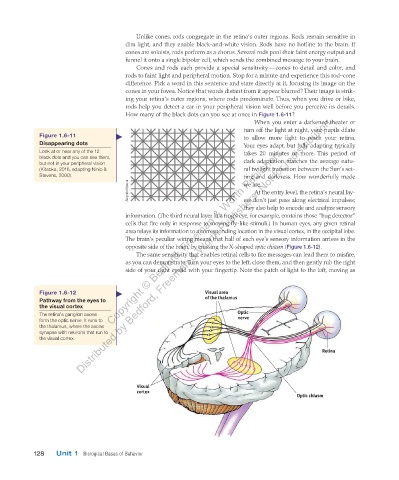
When you enter a darkened theater or
turn off the light at night, your pupils dilate
Figure 1.6-11 to allow more light to reach your retina.
Disappearing dots Your eyes adapt, but fully adapting typically
Copyright © Bedford, Freeman & Worth Publishers.
Look at or near any of the 12 takes 20 minutes or more. This period of
black dots and you can see them, dark adaptation matches the average natu-
but not in your peripheral vision
(Kitaoka, 2016, adapting Ninio & ral twilight transition between the Sun’s set-
Stevens, 2000). ting and darkness. How wonderfully made
Akiyoshi Kitaoka we are.
At the entry level, the retina’s neural lay-
ers don’t just pass along electrical impulses;
they also help to encode and analyze sensory
information. (The third neural layer in a frog’s eye, for example, contains those “bug detector”
cells that fire only in response to moving fly-like stimuli.) In human eyes, any given retinal
area relays its information to a corresponding location in the visual cortex, in the occipital lobe.
The brain’s peculiar wiring means that half of each eye’s sensory information arrives in the
opposite side of the brain, by crossing the X-shaped optic chiasm (Figure 1.6-12).
The same sensitivity that enables retinal cells to fire messages can lead them to misfire,
as you can demonstrate. Turn your eyes to the left, close them, and then gently rub the right
side of your right eyelid with your fingertip. Note the patch of light to the left, moving as
Figure 1.6-12 Visual area
Pathway from the eyes to of the thalamus
the visual cortex
The retina’s ganglion axons Optic
form the optic nerve. It runs to nerve
the thalamus, where the axons
synapse with neurons that run to
the visual cortex.
Retina
Visual
cortex
Optic chiasm
128 Unit 1 Biological Bases of Behavior
03_myersAPpsychology4e_28116_ch01_002_163.indd 128 15/12/23 9:25 AM