
Page 130 - 2024-bfw-MyersAP4e
P. 130
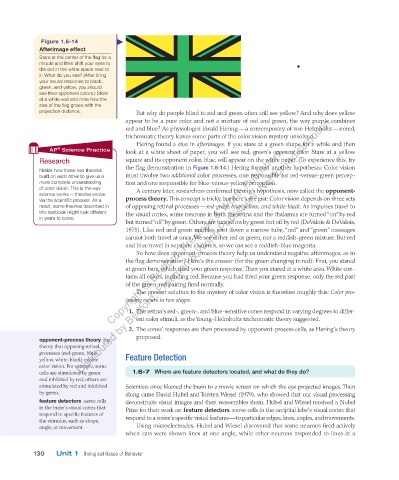
Figure   1.6-14
                    Afterimage effect
                   Stare at the center of the flag for a
                   minute and then shift your eyes to
                   the dot in the white space next to
                   it. What do you see? (After tiring
                   your neural response to black,
                   green, and yellow, you should
                   see their opponent colors.) Stare
                   at a white wall and note how the
                   size of the flag grows with the
                   projection distance.              But why do people blind to red and green often still see yellow? And why does yellow
                                 Distributed by Bedford, Freeman & Worth Publishers. Not for redistribution.
                                                appear to be a pure color and not a mixture of red and green, the way purple combines
                                                red and blue? As physiologist Ewald Hering — a contemporary of von Helmholtz — noted,
                                                trichromatic theory leaves some parts of the color vision mystery unsolved.
                                                     Hering found a clue in  afterimages.  If you stare at a green shape for a while and then
                                           Copyright © Bedford, Freeman & Worth Publishers.
                         ®
                      AP  Science Practice      look at a white sheet of paper, you will see red, green’s  opponent color. Stare at a yellow

                             Research           square and its opponent color, blue, will appear on the white paper. (To experience this, try

                                                the flag demonstration in  Figure 1.6-14. ) Hering formed another hypothesis: Color vision
                     Notice how these two theories
                   build on each other to give us a   must involve two  additional  color processes, one responsible for red-versus-green percep-
                   more complete understanding   tion and one responsible for blue-versus-yellow perception.
                   of color vision. This is the way         A century later, researchers confirmed Hering’s hypothesis, now called the    opponent-

                   science works — theories evolve

                   via the scientific process. As a   process theory  . This concept is tricky, but here’s the gist: Color vision depends on three sets

                   result, some theories described in   of opposing retinal processes —  red-green, blue-yellow,  and   white-black. As impulses travel to
                   this textbook might look different   the visual cortex, some neurons in both the retina and the thalamus are turned “on” by red
                   in years to come.
                                                but turned “off” by green. Others are turned on by green but off by red ( DeValois & DeValois,
                                                1975 ). Like red and green marbles sent down a narrow tube, “red” and “green” messages
                                                cannot both travel at once. We see either red or green, not a reddish-green mixture. But red
                                                and blue travel in separate channels, so we  can  see a reddish-blue magenta.
                                                         So how does opponent-process theory help us understand negative afterimages, as in
                                                the flag demonstration? Here’s the answer (for the green changing to red): First, you stared
                                                at green bars, which tired your green response. Then you stared at a white area. White con-
                                                tains all colors, including red. Because you had tired your green response, only the red part
                                                of the green-red pairing fired normally.
                                                     The present solution to the mystery of color vision is therefore roughly this:  Color pro-
                                                cessing occurs in two stages.

                                                     1.   The retina’s red-, green-, and blue-sensitive cones respond in varying degrees to differ-
                                                   ent color stimuli, as the Young–Helmholtz trichromatic theory suggested.

                                                    2.   The cones’ responses are then processed by opponent-process cells, as Hering’s theory
                                                   proposed.
                         opponent-process theory       the
                   theory that opposing retinal
                   processes (red-green, blue-
                   yellow, white-black) enable      Feature Detection
                   color vision. For example, some

                                                              e ar

                                                                 e featur
                                                                        e detectors located, and what do they do?


                                                              1.6-7
                                                          Wher
                   cells are stimulated by green               1.6-7    Where are feature detectors located, and what do they do?
                   and inhibited by red; others are
                   stimulated by red and inhibited     Scientists once likened the brain to a movie screen on which the eye projected images. Then
                   by green.                    along came David Hubel and Torsten Wiesel (1979), who showed that our visual processing
                      feature detectors       nerve cells   deconstructs visual images and then reassembles them. Hubel and Wiesel received a Nobel
                   in the brain’s visual cortex that   Prize for their work on   feature detectors   nerve cells in the occipital lobe’s visual cortex that
                                                                                   ,


                   respond to specific features of   respond to a scene’s specific visual features — to particular edges, lines, angles, and movements.
                   the stimulus, such as shape,
                   angle, or movement.               Using microelectrodes, Hubel and Wiesel discovered that some neurons fired actively
                                                when cats were shown lines at one angle, while other neurons responded to lines at a
                 130   Unit 1  Biological Bases of Behavior

          03_myersAPpsychology4e_28116_ch01_002_163.indd   130                                                                  15/12/23   9:25 AM