
Page 131 - 2024-bfw-MyersAP4e
P. 131
Module 1.6b
different angle. They surmised that these specialized neurons, now known as feature detec-
tors, receive information from individual ganglion cells in the retina. Feature detectors
pass this specific information to other cortical areas, where teams of cells (supercell clusters)
respond to more complex patterns.
For biologically important objects and events, monkey brains (and surely ours as well)
have a “vast visual encyclopedia” distributed in the form of specialized cells (Perrett et al.,
1990, 1992, 1994). These cells respond to one type of stimulus, such as a specific gaze, head
angle, posture, or body movement. Other supercell clusters integrate this information
and fire only when the cues collectively indicate the direction of someone’s attention and
approach. This instant analysis, which aided our ancestors’ survival, also helps a hockey
player anticipate where to shoot the puck, and a driver to anticipate a pedestrian’s next
Distributed by Bedford, Freeman & Worth Publishers. Not for redistribution.
movement.
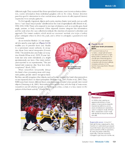
As we noted in Module 1.4, one tempo-
ral lobe area by your right ear (Figure 1.6-15) Figure 1.6-15
Copyright © Bedford, Freeman & Worth Publishers.
enables you to perceive faces and, thanks How your brain processes
to a specialized neural network, to recog- faces
nize them from varied viewpoints (Connor, In social animals such as
2010). This fusiform face area helps us recog- humans, a large right temporal
nize friends (Wiese et al., 2019). If your fusi- lobe area (shown here in a right-
form face area were stimulated, you might facing brain) is dedicated to the
crucial task of face recognition.
spontaneously see faces. One study partici- Viewing famous people’s faces,
pant reported to an experimenter, “You just compared with famous buildings,
turned into someone else. Your face meta- increases activation in this
fusiform face area (Gorno-Tempini
morphosed” (Koch, 2015). Face recognition area & Price, 2001).
When researchers temporarily disrupt (fusiform face area)
the brain’s face-processing areas with mag-
netic pulses, people cannot recognize faces.
But they can still recognize other objects, such as houses, because the brain’s face perception
occurs separately from its object perception (McKone et al., 2007; Pitcher et al., 2007). Thus,
fMRI scans have shown different brain areas becoming activated when people view varied
objects (Downing et al., 2001). Brain activity is so specific that, with the help of brain scans,
researchers can tell whether people are “looking at a shoe, a chair, or a face, based on the
pattern of their brain activity” (Haxby, 2001).
®
AP Science Practice
Research
An fMRI is often used as an
operational definition of brain
activity in neuroscience research.
Operational definitions are the
exact procedures (or operations)
used in a research study. Without
them, researchers couldn’t repli-
cate studies to ensure the results
are valid.
Supercells score In this 2017
Patrick McDermott/Getty Images Ovechkin (in red) instantly processed
National Hockey League game, Alex
visual information about the positions
and movements of three opponents.
By using his pattern-detecting
supercells, Ovechkin somehow
managed to get the puck into the net.
Sensation: Vision Module 1.6b 131
03_myersAPpsychology4e_28116_ch01_002_163.indd 131 15/12/23 9:25 AM