
Page 65 - 2024-bfw-MyersAP4e
P. 65
Module 1.4b
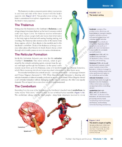
The brainstem is also a crossover point, where most nerves
to and from each side of the brain connect with the body’s FIGURE 1.4-7
opposite side (Figure 1.4-7). This peculiar cross- wiring — the The body’s wiring
brain’s contralateral hemispheric organization — is but one of
the brain’s many surprises.
The Thalamus
Sitting atop the brainstem is the forebrain’s thalamus, a pair forebrain consists of the
of egg-shaped structures that act as the brain’s sensory control cerebral cortex, thalamus, and
center (see Figure 1.4-6). The thalamus receives information hypothalamus; manages complex
Distributed by Bedford, Freeman & Worth Publishers. Not for redistribution.
from all the senses except smell, and routes that information cognitive activities, sensory
to the brain regions that deal with seeing, hearing, tasting, and and associative functions, and
voluntary motor activities.
touching. The thalamus also receives some of the replies from
those regions, which it then directs to the medulla and to the brainstem the central core
of the brain, beginning where
Copyright © Bedford, Freeman & Worth Publishers.
hindbrain’s cerebellum. Think of the thalamus as being to sen- the spinal cord swells as it
sory information what Seoul is to South Korea’s trains: a hub enters the skull; the brainstem
through which traffic passes en route to various destinations. is responsible for automatic
survival functions.
The Reticular Formation medulla [muh-DUL-uh] the
hindbrain structure that is
Inside the brainstem, between your ears, lies the reticular the brainstem’s base; controls
(“netlike”) formation. This nerve network, which is gov- heartbeat and breathing.
erned by the reticular activating system, extends from the spi- thalamus [THAL-uh-muss]
nal cord right up through the thalamus. As the spinal cord’s the forebrain’s sensory control
sensory input flows up to the thalamus, some of it travels through the reticular formation, center, located on top of the
which filters incoming stimuli and relays important information to other brain areas. brainstem; it directs messages to
the sensory receiving areas in the
The reticular formation also controls arousal — our state of alertness — as Giuseppe Moruzzi cortex and transmits replies to
and Horace Magoun discovered in 1949. When they electrically stimulated a sleeping cat’s the cerebellum and medulla.
reticular formation, it almost instantly produced an awake, alert animal. When Magoun severed reticular formation a nerve
a cat’s reticular formation without damaging nearby sensory pathways, the effect was equally network that travels through the
dramatic: The cat lapsed into a coma from which it never awakened. brainstem into the thalamus;
it filters information and plays
The Cerebellum an important role in controlling
arousal.
Extending from the rear of the brainstem is the hindbrain’s baseball-sized cerebellum; its cerebellum [sehr-uh-
name means “little brain,” which is what its two wrinkled halves resemble (Figure 1.4-8). BELL-um] the hindbrain’s
The cerebellum (along with the basal ganglia — deep brain structures involved in motor “little brain” at the rear of the
brainstem; its functions include
processing sensory input,
coordinating movement output
and balance, and enabling
nonverbal learning and memory.
Figure 1.4-8
Tony Quinn/ZUMA Press/Newscom voluntary movements, as when
The brain’s organ of agility
the cerebellum coordinates our
Cerebellum Hanging at the back of the brain,
Spinal cord soccer player Mallory Pugh
controls the ball.
The Brain: Brain Regions and Structures Module 1.4b 65
03_myersAPpsychology4e_28116_ch01_002_163.indd 65 15/12/23 9:23 AM