
Page 60 - 2024-bfw-MyersAP4e
P. 60
blood rushing to the back of the brain, which processes visual infor-
mation. Another tool, functional near-infrared spectroscopy (fNIRS),
shines infrared light on blood molecules to identify brain activity.
Mark Straccia/UCLA Social Cognitive Neuroscience Laboratory (Burns et al., 2019; Perdue et al., 2019). Table 1.4-1 compares imag-
The fNIRS equipment can fit in a large backpack, enabling research-
ers to study the biology of mind in difficult-to-reach populations
ing techniques.
Such snapshots of the brain’s activity provide new insights
into how the brain divides its labor and reacts to changing needs.
A mountain of recent fMRI studies has revealed which brain areas
are most active when people feel pain or rejection, listen to angry
Distributed by Bedford, Freeman & Worth Publishers. Not for redistribution.
aroused. The technology enables a very basic sort of mind reading.
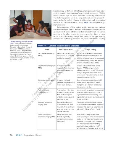
Understanding the non-WEIRD voices, think about scary things, feel happy, or become sexually
Copyright © Bedford, Freeman & Worth Publishers.
brain Most neuroscience research
studies people from Western, TABLE 1.4-1 Common Types of Neural Measures
Educated, Industrialized, Rich, and
Democratic (WEIRD) populations Name How Does It Work? Sample Finding
(Falk et al., 2013). Using functional
near-infrared spectroscopy (fNIRS), Electroencephalography Electrodes placed on the Symptoms of depression and anxiety
the researchers shown here were
able to identify brain areas involved in (EEG) scalp measure electrical correlate with increased activity in the
persuasion among a Jordanian sample activity in neurons. right frontal lobe, a brain area associated
(Burns et al., 2019).
with behavioral withdrawal and negative
emotion (Thibodeau et al., 2006).
Magnetoencephalography A head coil records Soldiers with posttraumatic stress
(MEG) magnetic fields from the disorder (PTSD), compared with
brain’s natural electrical soldiers who do not have PTSD, show
currents. stronger magnetic fields in the visual
cortex when they view trauma-related
images (Todd et al., 2015).
Computed tomography X-rays of the head Children’s brain injuries, shown in CT
(CT) generate images that may scans, predict impairments in their
locate brain damage. intelligence and memory processing
(Königs et al., 2017).
Positron emission Tracks where in the brain Monkeys with an anxious temperament
tomography (PET) a temporarily radioactive have brains that use more glucose in
form of glucose goes regions related to fear, memory, and
while the person given it expectations of reward and punishment
performs a task. (Fox et al., 2015).
Magnetic resonance People sit or lie down People with a history of violence tend
imaging (MRI) in a chamber that uses to have smaller frontal lobes, especially
magnetic fields and radio in regions that aid moral judgment and
waves to provide a map self-control (Glenn & Raine, 2014).
of brain structure.
Functional magnetic Measures blood flow Years after surviving a near plane
resonance imaging (fMRI) to brain regions by crash, passengers who viewed material
comparing continuous related to their trauma showed greater
MRI scans. activation in the brain’s fear, memory,
and visual centers than when they
watched footage related to the 9/11
terrorist attacks (Palombo et al., 2015).
60 Unit 1 Biological Bases of Behavior
03_myersAPpsychology4e_28116_ch01_002_163.indd 60 29/12/23 12:14 PM